Background on bone and femur:
Bone is a hard tissue which is robust against physiological loads.
The position of the bone and its function within the skeletal frame limits its range of motion through linkages between bones, joints and muscle attachment.
Muscles attach to bones through tendons.
Femur is indispensable for locomotion and load-bearing functions within a large range of motion under potentially high load magnitudes and impacts. The ability of the femur to withstand these loads is achieved through adaptive processes that modulate its morphology and composition.
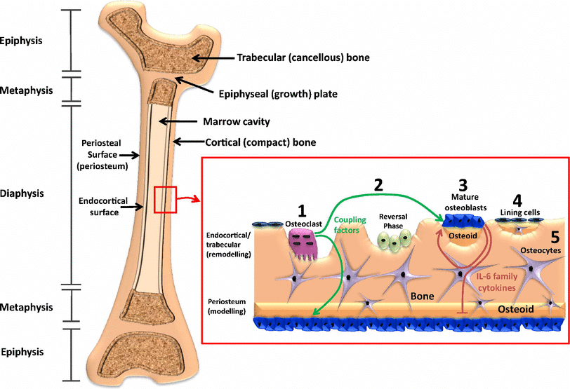
Morphology involves examining bones at different levels, from the macroscopic features visible to the naked eye to the microscopic details observed at the cellular and tissue levels.
The cortical geometry at the proximal femur consequently reflects the most robust construction adapted for the specific loading it habitually experiences e.g. locomotion and physical activity. Thus, consistent loading in specific directions, such as physical training of athletes over long period of time, can induce corresponding local adaptations in the cortical geometry.
While loading stimuli have the most pronounced effect in adolescence, their effectiveness in stimulating an adaptation decreases after bone reaches maturity. However, it is known that the exercise-induced bone thickening during growth occurs through new bone formation on the periosteal bone surface, while age-related bone loss takes place at the endocortical bone surface. Thus, beneficial geometric adaptations accrued during adolescence and young adulthood may have lasting benefits in senescence despite associated bone loss.
Understanding the mechanisms that modulate specific adaptations at bone regions susceptible to fractures can be used as paradigms for preventing age-related degradation of bone robustness, or alternatively, increased bone fragility. Also, establishing morphological features that result from specific loading patterns may help in recreating the activity of past population in the field of archaeological anthropology.
Literature review:
Studies exploring the spatial heterogeneity in the adaptive response to physical loading have traditionally analysed bone cross-sections with respect to mechanically relevant (morphometric) features. (Morphometric features include measurements related to bone geometry, such as cortical thickness, bone volume, and various geometric parameters.)
To increase detectability of this heterogeneity it is necessary to increase the spatial resolution of analyses. Within femoral neck cross-sections this has been achieved by partitioning into quadrants, octants or higher angular divisions defined with respect to anatomical directions.
Access to 3D tomographic data has extended the potential for such detailed analyses. Concepts of computational anatomy and statistical parametric mapping make it possible to investigate the distribution of relevant parameters over 3D regions and access their statistical significance.
In large sample studies, establishing anatomical correspondance across samples is a prerequisite. Often noise and varying, or even incomplete, anatomical extents in the data can compromise the consistency in the shape registration process. (It may involve creating a spatial mapping that allows for a meaningful comparison of anatomical shapes and structures, facilitating group-level analyses, statistical comparisons, and population-based studies.) Thus methods that can provide computational registration across multiple surface instances can expand the tool-set beyond the commonly used iterative closest point (ICP) based methods.
Ricci-flow:
This study introduces a novel application of the conformal mapping method for establishing correspondance between proximal femur instances. The method treats the surface as a differentiable manifold and converts the 3D registration problem into 2D. This dimension reduction is achieved by a parametrisation procedure where the surface is conformally mapped to the planar domain using discrete Ricci-flow. The advantages of this method in shape registration and indexing have been exhibited for anatomical objects such as brain and colon.
Dataset:
The present datasets consists of young adult females whose consistent and vigorous training regimen represented habitual activity. Geometric morphology was quantified in terms of the spatial distribution of cortical thickness. Adaptation was consequently defined spatially as regions exhibiting statistically significant differences from the controls.
The recent finite element (FE) study conducted by Abe and colleagues simulated a ‘supra-physiological’ loading caused by sideways falling, to assess fracture risk of proximal femur. Utilizing the results of this simulation, we illustrated the influence of adapted geometry in lowering fall-induced stresses in the femoral neck region.
The data consisted of tomographic MRI images of the proximal femur of 111 participants:
- 91 female athletes
- 20 physically active women serving as a control group
The MRI protocol was based on axis T1-weighted gradient echo VIBE examination.
The athletes were categorized into mutually exclusive exercise loading groups by their characteristics loading patterns in their respective sports according to our standard procedure.
- high impact (HI) group associated with maximal vertical jumps and high impacts (High jumpers and long jumpers, N=17)
- odd impact (OI) group associated with rapid acceleration and deceleration as well as moderate to high impacts and bending forces from varying directions (Squash and soccer players, N=19)
- high magnitude (HM) group associated with movements with coordinated high muscle force production and low rate (Powerlifters, N=17)
- repetitive impact (RI) group associated with highly repetitive weight bearing impacts and bending force (Endurance runners, N=18)
- repetitive non impact (RNI) group associated with highly repetitive movements lacking ground impacts (Swimmers, N=18)
The anatomy of interest - proximal femur cortical geometry - was manually segmented from the MRI data of each participant. Apparently, the limited in-plane resolution of the native MR images compromises reliable inferences at locations with very thin cortices such as the femoral head and some regions of the femoral neck and the trochanters. However, it has been shown that MR imaged cross-sections of the femoral neck are sufficiently precise and accurate for study purposes.
Extracted 3D cortical geometry to 2D mesh:
To extract the proximal femur cortical geometry, the image data of each participant was manually segmented by delineating periosteal and endosteal cortical surfaces. Thereafter the segmented geometry was converted into a volume mesh (10-node tetrahedral elements) for constructing a finite element (FE) model. The nodes from the periosteal and endosteal cortical surfaces of the volume mesh were extracted, along with the maximum and minimum principal stain values from the above-noted simulation results. (This mesh is a discretized representation of the anatomical structure, with tetrahedral elements serving as the basic building blocks.) The selected nodes were used to reconstruct the inner and outer triangular surface meshes of the cortical bone in MeshLab. The nodes in the point cloud () were first down-sampled to using the Poisson disk sampling module and a surface reconstructed using the ball-pivoting module. After checking for and cleaning any major errors (e.g. intersections, face flips, duplicates, hoes) in the surfaces, they were remeshed to improve the quality (aspect ratio < 20) of the mesh in Avizo.
Planar parametrisation: Discrete Ricci-flow:
To analyse and contrast the group-wise morphology of the proximal femurs, it was essential to establish correspondence between all individual surface meshes. The approach used in this work transforms the 3D surfaces into 2D (planar domain) using an angle preserving conformal method based on Ricci-flow. This approach leverages the methodological advantages of invariance towards rigid motion, scale and isometric defomration. The residual deformations such as large non-isotropic deformations can then subsequently be accounted for in the 2D domain which is relatively a simpler task.
The discrete representation of the femur surfaces embedded in the Euclidean space allows them to be treated as differentiable manifolds (2-manifold). Any surface in Euclidean space is a Riemannian surface with an inherent metric called Riemannian metric and a conformal structure. The Uniformisation theorem states that the Riemannian metric can be deformed to admit uniform curvature over the surface: . Thus any closed Riemannian surface can be conformally mapped to one of these fundamental surfaces: unit sphere (+1), Euclidean plane (0) or hyperbolic plane (-1). The embedding in the relevant domain, referred to as parametrisation, reflects an angle preserving (conformal) transformation. Ricci-flow is a robust curvature flow method introduced by Hamilton that evolves the metric towards uniformisation as a heat diffusion process. It is a powerful method that provides the flexibility to design the final metric based on user-defined distribution of target Gaussian curvature. However, the total curvature is determined by the topology of the surface (S) according to Gauss-Bonnet theorem: . For an open orientable surface of genus () and number of boundaries , the Euler number is given by . The genus of a closed surface can be intuitively thought of as the number of handles contained. The proximal femur data presented challenges that were addressed by tailoring specific solutions.
The foremost challenge was the lack of anatomical correspondance at the distal boundary (femur diaphysis) of he surfaces across subjects because of different field of views. This inconsistency of the boundary made it unreliable reference for the subsequent correspondence detection. Thus appropriate references were derived from surface features through two step parameterisation procedure. The first step was devised to detect shape features to introduce a consistently defined reference feature for each mesh. The second step was devised to produce a mapping in a common coordinate space using the reference points for alignment.
The topology of outer cortical surface () is conformally equivalent to disk . Ricci-flow mapping was performed by assigning zero target curvature to all interior points and leaving the boundary node metric unchanged i.e a free boundary condition. The resulting conformal factor distribution was used to investigate detectable surface features. Two prominent features processes were identified from the disk parametrisation - the femoral head (FH) and the greater trochanter (GT). The two features can be seen as peaks whose representative centers were designated as feature points. These feature points were used consistently across all participants to introduce a stable reference in the form of an inter-feature geodesic. This was achieved by inserting a boundary (mesh slit) along the inter-feature geodesic path in the parametrised disk (i.e. straight line) and reflected in the native 3D surface. This modified surface () is conformally equivalent to Euclidean annulus. The mapping was performed by assigning a target curvature of 0 to all nodes. A cut graph between the GT point and the distal boundary lay on the imaginary axis with distal boundary parallel to it and incident on the negative real axis. Finally the mesh was resized such that the inter-feature boundary (on the imaginary axis) was scaled to . The annulus was produced through an exponential map of the complex coordinates.